具体描述
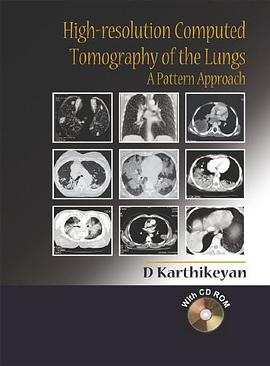
The advent of chest CT with high-resolution techniques has changed dramatically the understanding of pulmonary diseases. Conditions previously hard to distinguish using traditional film radiography, particularly the diffuse lung diseases, can now be diagnosed rapidly and the extent of the disease process identified. Designed for easy reference in the clinical setting, this highly illustrated 'text and atlas' is a comprehensive but practical guide to performing and interpreting CT imaging studies of the chest. Opening with a review of the fundamentals of high-resolution CT in relation to lung chest anatomy, the second section forming the bulk of the book is a case-based review of both focal and diffuse lung diseases, describing the features of those diseases as visualised using CT and related differential diagnoses where relevant. The book concludes with an extensive appendix of useful information relating to chest imaging, including key facts about all the commonly encountered pathologic entities and protocols that can be referred to in the clinical setting. The book is accompanied by a CD containing all the images from the book with a presentation on pattern approach that can be used for teaching purposes as well as self-assement.
作者简介
目录信息
读后感
评分
评分
评分
评分
用户评价
作为一名影像学爱好者,我一直对医学影像的精细化处理非常着迷。尤其是肺部CT,那些微小的结构变化常常隐藏着疾病的关键信息。《High-resolution Computed Tomography of the Lungs》这个书名,立刻引起了我的注意。我猜这本书可能会深入探讨高分辨率CT成像的技术原理,比如不同探测器类型、扫描层数、以及重建算法如何影响图像的细节表现。我特别想了解,在保持低辐射剂量的同时,如何才能获得高质量的高分辨率图像,以及如何利用后处理技术,例如多平面重建(MPR)、三维重建(3D VR)、以及钙化积分(CAL)等,来更全面地展示肺部结构。我期待书中能有关于如何识别和鉴别肺部微小结节,包括其边缘、密度、内部结构等特征的详细指导。此外,对于肺部感染、肺栓塞、肺纤维化等常见疾病,书中是否能提供一些高分辨率CT下特有的、能够帮助我们进行早期诊断和精准分型的影像学表现。我还希望书中能包含一些关于CT成像辐射剂量的管理和优化策略,以及如何在保证成像质量的前提下,最大限度地降低患者的辐射暴露。
评分我是一名肺科医生,对肺部影像学一直抱有极大的兴趣。在我看来,影像学是诊断和治疗肺部疾病的基石。每次看到那些高清晰度的肺部CT图像,我都感觉自己离疾病的真相又近了一步。《High-resolution Computed Tomography of the Lungs》这个书名,让我充满了期待。我希望这本书能够详细介绍高分辨率CT在肺部疾病诊断中的具体应用,比如如何利用它来更早地发现和评估肺结节,如何区分良恶性结节,以及如何监测结节的生长和变化。我还想知道,书中是否会涵盖高分辨率CT在间质性肺病、肺气肿、支气管扩张症等疾病中的详细表现,以及如何通过图像特征来判断疾病的严重程度和预后。此外,对于一些罕见的肺部疾病,例如肺血管炎、淋巴瘤等,书中是否能提供一些有价值的影像学线索,帮助我们进行早期诊断。我更希望这本书能够提供大量的图例,并且对每张图例进行深入的分析和解读,让我能够从中学习到如何像专家一样去阅读和理解肺部的高分辨率CT图像。
评分这本书名《High-resolution Computed Tomography of the Lungs》一听就相当专业,对于我这种临床经验尚浅的医生来说,简直是如获至宝。我一直觉得CT成像的细节处理是诊断的关键,尤其是在肺部疾病领域,那些微小的病灶往往决定了治疗的方向和预后。看到这个书名,我立刻联想到无数次在阅片室里纠结于那些模糊不清的影迹,希望能有更清晰、更直观的图像来指导我的判断。这本书的出现,仿佛是一盏明灯,照亮了我探索肺部CT成像奥秘的道路。我迫不及待地想知道,它究竟是如何做到“High-resolution”的,是通过新型的扫描技术?还是先进的图像后处理算法?我特别期待书中能够详细解析这些技术细节,比如不同层厚、不同重建算法对病灶显示的影响,以及如何通过优化扫描参数来最大化信息量。另外,对于那些常见的、但又容易被忽略的微小征象,比如早期肺栓塞的微小实变、早期间质性肺病的磨玻璃影,这本书是否能提供更精细的鉴别诊断方法,从而帮助我们更早、更准确地发现和诊断这些疾病?我希望书中不仅有理论介绍,更能提供大量的典型病例图片,并配以详细的解读,让我能将书本知识与实际阅片相结合,真正提升自己的诊断能力。
评分我是一名资深的影像科医师,在肺部CT诊断领域已经工作多年。尽管经验丰富,但我始终认为学习永无止境,尤其是在快速发展的医学影像技术面前。这本书《High-resolution Computed Tomography of the Lungs》这个书名,让我眼前一亮。我猜测这本书的作者一定是对肺部高分辨率CT成像有着深入的研究和独到的见解。我特别感兴趣的是,书中是否会对当前主流的高分辨率CT技术,例如薄层扫描、容积扫描、以及各种后处理算法(如最大密度投影、最小密度投影、曲面重构等)进行系统性的梳理和评价。我希望它能提供一些关于如何根据不同疾病特点,选择最适合的高分辨率成像参数和后处理方法的指导。此外,对于那些疑难病例,例如早期弥漫性肺部疾病、肺部感染的复杂表现、或者肿瘤的微小转移灶,书中是否能提供一些鉴别诊断的线索和技巧,尤其是在高分辨率CT图像上的细微特征解读。我还想知道,书中是否会涉及到一些人工智能辅助诊断在高分辨率肺部CT成像中的应用前景和技术进展,以及如何利用这些技术来提高诊断的效率和准确性。
评分我是一名影像科的住院医生,目前正在积极学习胸部CT的判读。我之前接触过一些CT的教材,但总觉得在高分辨率成像方面,市面上能找到的资料深度和广度都有限。尤其是在处理复杂的肺部疾病时,比如一些少见病或者早期病变,分辨率不足就成为一个很大的障碍。这本书《High-resolution Computed Tomography of the Lungs》这个书名,立刻就吸引了我。我猜测它可能会涵盖很多关于低剂量CT、双能量CT在高分辨率成像中的应用,以及如何利用最新的AI技术来增强图像质量和辅助诊断。我非常好奇书中是否会深入探讨如何识别和描述那些非常细微的肺部结构变化,比如早期肺气肿的肺泡壁增厚、细小的支气管扩张、或者是早期结节的形态学特点。还有,对于肺部淋巴管的显示,以及如何通过高分辨率CT来评估肺栓塞的严重程度和慢性肺栓塞的征象,我都希望能在这本书里找到答案。另外,我特别期待书中能有关于不同制造商的CT设备在高分辨率成像方面的对比分析,以及不同扫描和重建策略对不同类型肺部病变成像效果的影响。这种接地气的实用信息,对于我们在日常工作中选择最优的成像方案非常有帮助。
评分 评分 评分 评分 评分相关图书
本站所有内容均为互联网搜索引擎提供的公开搜索信息,本站不存储任何数据与内容,任何内容与数据均与本站无关,如有需要请联系相关搜索引擎包括但不限于百度,google,bing,sogou 等
© 2026 qciss.net All Rights Reserved. 小哈图书下载中心 版权所有


